


 Foundational characteristics of cancer include proliferation, angiogenesis, migration, evasion of apoptosis, and cellular immortality. Find key markers for these cellular processes and antibodies to detect them.
 The SUMOplot™ Analysis Program predicts and scores sumoylation sites in your protein. SUMOylation is a post-translational modification involved in various cellular processes, such as nuclear-cytosolic transport, transcriptional regulation, apoptosis, protein stability, response to stress, and progression through the cell cycle.
 The Autophagy Receptor Motif Plotter predicts and scores autophagy receptor binding sites in your protein. Identifying proteins connected to this pathway is critical to understanding the role of autophagy in physiological as well as pathological processes such as development, differentiation, neurodegenerative diseases, stress, infection, and cancer.
> home > Products > Primary Antibodies > Antibody Collections > Recombinant Antibodies > Anti-ANGPTL4 Reference Antibody (Nanyang Tech.U. patent anti-ANGPTL4)
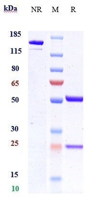
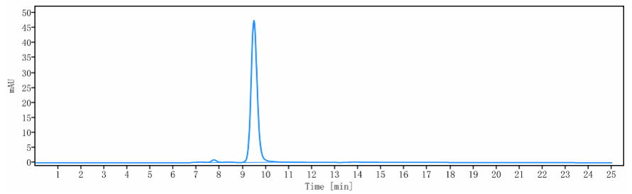
Anti-ANGPTL4 Reference Antibody (Nanyang Tech.U. patent anti-ANGPTL4)
Recombinant Antibody
- SPECIFICATION
- CITATIONS
- PROTOCOLS
- BACKGROUND

Application
| FC, Kinetics, Animal Model |
|---|---|
| Primary Accession | Q9BY76 |
| Reactivity | Human |
| Clonality | Monoclonal |
| Isotype | IgG1 |
| Calculated MW | 150 KDa |
| Target/Specificity | ANGPTL4 |
|---|---|
| Endotoxin | < 0.001EU/ µg,determined by LAL method. |
| Conjugation | Unconjugated |
| Expression system | CHO Cell |
| Format | Purified monoclonal antibody supplied in PBS, pH6.0, without preservative.This antibody is purified through a protein A column. |
| Name | ANGPTL4 |
|---|---|
| Synonyms | ARP4, HFARP, PGAR {ECO:0000303|PubMed:10 |
| Function | Mediates inactivation of the lipoprotein lipase LPL, and thereby plays a role in the regulation of triglyceride clearance from the blood serum and in lipid metabolism (PubMed:19270337, PubMed:21398697, PubMed:27929370, PubMed:29899144). May also play a role in regulating glucose homeostasis and insulin sensitivity (Probable). Inhibits proliferation, migration, and tubule formation of endothelial cells and reduces vascular leakage (PubMed:14583458, PubMed:17068295). Upon heterologous expression, inhibits the adhesion of endothelial cell to the extracellular matrix (ECM), and inhibits the reorganization of the actin cytoskeleton, formation of actin stress fibers and focal adhesions in endothelial cells that have adhered to ANGPTL4-containing ECM (in vitro) (PubMed:17068295). Depending on context, may modulate tumor-related angiogenesis (By similarity). |
| Cellular Location | Secreted. Secreted, extracellular space, extracellular matrix. Note=The unprocessed form interacts with the extracellular matrix (PubMed:17068295, PubMed:21398697). This may constitute a dynamic reservoir, a regulatory mechanism of the bioavailability of ANGPTL4 (Probable). |
| Tissue Location | Detected in blood plasma (at protein level) (PubMed:29899519). Detected in liver (PubMed:10698685). Detected in white fat tissue and placenta (PubMed:10866690). Expressed at high levels in the placenta, heart, liver, muscle, pancreas and lung but expressed poorly in the brain and kidney. |
Research Areas
Citations (0)

Thousands of laboratories across the world have published research that depended on the performance of antibodies from Abcepta to advance their research. Check out links to articles that cite our products in major peer-reviewed journals, organized by research category.
Submit your citation using an Abcepta antibody to
info@abcepta.com, and receive a free "I Love Antibodies" mug.
info@abcepta.com, and receive a free "I Love Antibodies" mug.

Application Protocols
Provided below are standard protocols that you may find useful for product applications.
Abcepta welcomes feedback from its customers.
If you have used an Abcepta product and would like to share how it has performed, please click on the "Submit Review" button and provide the requested information. Our staff will examine and post your review and contact you if needed.
If you have any additional inquiries please email technical services at tech@abcepta.com.
$ 385.00
Cat# APR10783
Ordering Information
United States
AlbaniaAustraliaAustriaBelgiumBosnia & HerzegovinaBrazilBulgariaCanadaCentral AmericaChinaCroatiaCyprusCzech RepublicDenmarkEstoniaFinlandFranceGermanyGreeceHong KongHungaryIcelandIndiaIndonesiaIrelandIsraelItalyJapanLatviaLithuaniaLuxembourgMacedoniaMalaysiaMaltaMexicoNetherlandsNew ZealandNorwayPakistanPolandPortugalRomaniaSerbiaSingaporeSlovakiaSloveniaSouth AfricaSouth KoreaSpainSwedenSwitzerlandTaiwanTurkeyUnited KingdomUnited StatesVietnamWorldwideOthers
USA Headquarters
(888) 735-7227 / (858) 622-0099 or (858) 875-1900
Other Products
Shipping Information
Domestic orders (in stock items)
Shipped out the same day. Orders placed after 1 PM (PST) will ship out the next business day.
International orders
Contact your local distributors
